Blood Vessels Labeled Diagram : Types Of Blood Vessels And Their Further Classifications Labeled Diagram Of A Red Blood Cell Transparent Png 1432x875 Free Download On Nicepng : Labeled diagram showing the structure of a blood vessel observe the blood vessels diagrams above, where you can see the structures of arteries and veins clearly labeled.
Blood Vessels Labeled Diagram : Types Of Blood Vessels And Their Further Classifications Labeled Diagram Of A Red Blood Cell Transparent Png 1432x875 Free Download On Nicepng : Labeled diagram showing the structure of a blood vessel observe the blood vessels diagrams above, where you can see the structures of arteries and veins clearly labeled.. Spend a while piecing these diagrams together in your mind, trying to link the labeled names with the functions you learned about in the video. We then simplified the anatomy of the heart even further with the below cartoon diagram and 2x2 table. In this image, you will find blood vessel and capillary diagram, arteriole, precapillary sphincter, metarteriole, arteriovenous anastomosis, capillary, venule, thoroughfare channel in it. All blood vessels are basically hollow tubes with an internal space, called a lumen, through which blood flows. Just need a glimpse, leave your valuable advice let us know , and subscribe us!
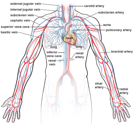
These vessels connect other organs in your body to your heart. Labeled diagram showing the structure of a blood vessel observe the blood vessels diagrams above, where you can see the structures of arteries and veins clearly labeled. Bulky middle tunic contains smooth muscle and elastin 3. A heart diagram labeled will provide plenty of information about the structure of your heart, including the wall of your heart. A primary purpose and significant role of the vasculature is its participation in oxygenating the body.
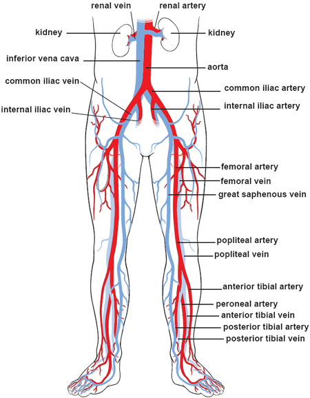
Illustrations Of The Blood Vessels from my.clevelandclinic.org Our latest youtube film is ready to run. The iliac, femoral, popliteal and tibial (calf) veins are the deep veins in the legs. Arterioles connect with even smaller blood vessels called capillaries. Anatomy of the heart and blood vessels. Arteries, arterioles, capillaries, venules and veins. Veins (in blue) are the blood vessels that return blood to the heart. It's coming from the lungs and going to the left atrium, so it's going to be the pulmonary vein woman. Learn vocabulary, terms, and more with flashcards, games, and other study tools.
10 photos of the the human blood vessels labeled.
Arteries, arterioles, capillaries, venules and veins. Blood vessels labeled diagram : We have an illustration of the cardiovascular system, and we're labeling the blood vessels. Learn even faster with this blood vessel anatomy study guide. The study of blood vessels is called angiology. The duodenum is supplied by the superior and inferior pancreaticoduodenal arteries, which are the branches of the gastroduodenal and superior mesenteric arteries, respectively. Blood vessel, a vessel in the human or animal body. Anatomy of the heart and main cardiac structures including the heart valves, chambers (atria and ventricles), and great vessels. A web of blood vessels—arteries, veins, and capillaries—circulate blood to organs. Blood vessels are the specially designed tubes that carry blood throughout the body. Tutorials and quizzes on the circulation of blood and the anatomy, structure, and physiology of blood vessels, using interactive animations and diagrams. Capillaries lead back to small vessels known as venules that flow into the larger veins and eventually back to the heart. A vein is a blood vessel that conducts blood toward the heart.
Anatomy of the heart and main cardiac structures including the heart valves, chambers (atria and ventricles), and great vessels. Includes an exercise, review worksheet, quiz, and model drawing of an anterior vi The walls of blood vessels differ depending on the type of vessel. Arteries transport blood away from the heart and branch into smaller vessels, forming arterioles. A vein is a blood vessel that conducts blood toward the heart.
Illustrations Of The Blood Vessels from my.clevelandclinic.org Hma practical 3 for monday july 23 and wednesday july 25. Arteries transport blood away from the heart and branch into smaller vessels, forming arterioles. All blood vessels are basically hollow tubes with an internal space, called a lumen, through which blood flows. The blood vessels are an intricate network of hollow tubular structures carrying blood throughout the body. Arterioles connect with even smaller blood vessels called capillaries. Huge collection, amazing choice, 100+ million high quality, affordable rf and rm images. The iliac, femoral, popliteal and tibial (calf) veins are the deep veins in the legs. In this image, you will find blood vessel and capillary diagram, arteriole, precapillary sphincter, metarteriole, arteriovenous anastomosis, capillary, venule, thoroughfare channel in it.
Learn even faster with this blood vessel anatomy study guide.
The blood vessels are an intricate network of hollow tubular structures carrying blood throughout the body. Blood vessels labeled diagram : Arteries (in red) are the blood vessels that deliver blood to the body. Arterioles connect with even smaller blood vessels called capillaries. The heart beats continuously, pumping the equivalent of more than 14,000 litres of blood every day through five main types of blood vessels: Its smooth surface decreases resistance to blood flow The walls of blood vessels differ depending on the type of vessel. Deep veins, located in the center of the leg near the leg bones, are enclosed by muscle. Blood vessel, a vessel in the human or animal body. 10 photos of the the human blood vessels labeled. Coronary circulation anatomical cross section diagram, labeled vector illustration scheme. Vessels of the small intestine are grouped by which segment they supply: Spend a while piecing these diagrams together in your mind, trying to link the labeled names with the functions you learned about in the video.
Arteries carry oxygenated blood except in case of the pulmonary artery. Deoxygenated blood from the peripheral veins is transported back to the heart from capillaries, to venules, to veins, to the right side of the heart, and then. A primary purpose and significant role of the vasculature is its participation in oxygenating the body. Learn even faster with this blood vessel anatomy study guide. Venous drainage occurs via the prepyloric.
Honors Anatomy Chapter 11 Unit 3 Blood Vessels And Circulation Diagram Quizlet from o.quizlet.com Use key choices to identify the blood vessel tunic described. The walls of blood vessels differ depending on the type of vessel. Hma practical 3 for monday july 23 and wednesday july 25. They transport blood cells, nutrients and oxygen and carry away carbon dioxide and waste materials from the tissues and organs. Bulky middle tunic contains smooth muscle and elastin 3. A heart diagram labeled will provide plenty of information about the structure of your heart, including the wall of your heart. Labeled diagram showing the structure of a blood vessel observe the blood vessels diagrams above, where you can see the structures of arteries and veins clearly labeled. Veins (in blue) are the blood vessels that return blood to the heart.
Arterioles distribute blood to capillary beds, the sites of exchange with the body tissues.
They also take waste and carbon dioxide away from the tissues. Its smooth surface decreases resistance to blood flow Start studying blood vessels labeling. The iliac, femoral, popliteal and tibial (calf) veins are the deep veins in the legs. From the center of the optic nerve radiates the major blood vessels of the retina. The heart is a muscular pump that pushes blood through blood vessels around the body. A vein is a blood vessel that conducts blood toward the heart. Deep veins, located in the center of the leg near the leg bones, are enclosed by muscle. Arteries transport blood away from the heart and branch into smaller vessels, forming arterioles. Blood vessels labeled diagram, blood vessels labeling exercises, cat blood vessels labeled, human anatomy blood vessels, human. 10 photos of the the human blood vessels labeled. Blood vessels labeled diagram, blood vessels labeling exercises, cat blood vessels labeled, human anatomy blood vessels, human blood vessels images, human blood vessels length, human blood vessels video, human body blood vessels, inner body, blood vessels labeled diagram, blood vessels. Capillaries lead back to small vessels known as venules that flow into the larger veins and eventually back to the heart.
Anatomy of blood vessels review sheet 32 261 microscopic structure of the blood vessels 1 blood vessels labeled. A primary purpose and significant role of the vasculature is its participation in oxygenating the body.


0 Komentar